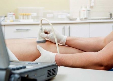
Ultrasound Musculoskeletal imaging of the musculoskeletal system is a powerful diagnostic tool used to assess and evaluate muscles, joints, tendons, ligaments, and other soft tissues. This non-invasive and radiation-free imaging technique provides real-time insights into the structure and function of the musculoskeletal system. Whether you’re dealing with sports injuries, chronic pain, or other conditions, musculoskeletal ultrasound offers precise diagnostic capabilities. For those in need of Musculoskeletal Ultrasound services in Pune, the Insight Imaging Clinic in Thergaon, Pune, led by Dr. Snehal Suryawanshi, is a trusted destination. Renowned as the Best Radiodiagnosis Clinic in Pune, the clinic is known for its cutting-edge technology and exceptional patient care.
What is Musculoskeletal Ultrasound?
Musculoskeletal ultrasound is a specialized imaging technique that uses high-frequency sound waves to produce images of the soft tissues and structures within the musculoskeletal system. Unlike traditional imaging methods, it allows for dynamic assessment, enabling healthcare providers to observe structures in motion and pinpoint abnormalities with precision.
What is Ultrasound Imaging of the Musculoskeletal System?
This imaging modality focuses on diagnosing and managing conditions affecting:
- Muscles: To detect tears, strains, or abnormalities.
- Tendons: For identifying inflammation, tears, or ruptures.
- Ligaments: To evaluate sprains or tears.
- Joints: To assess effusions, synovitis, or cartilage damage.
- Nerves: To detect entrapments or injuries.
Musculoskeletal ultrasound is particularly valuable for guiding minimally invasive procedures such as joint injections or biopsies.
What Are Some Common Uses of the Procedure?
Musculoskeletal ultrasound is used in a variety of scenarios, including:
- Sports Injuries: Diagnosing sprains, strains, or tendon tears in athletes.
- Arthritis: Evaluating joint inflammation and tracking disease progression.
- Bursitis and Tendinitis: Detecting inflammation in bursae or tendons.
- Carpal Tunnel Syndrome: Identifying nerve compression in the wrist.
- Cysts and Masses: Locating and assessing soft tissue lumps or cysts.
- Fractures: Visualizing small or hidden fractures.
What Does the Equipment Look Like?
The main components of musculoskeletal ultrasound equipment include:
Transducer (Probe):
A handheld device that emits high-frequency sound waves and captures the echoes reflected from tissues.
Ultrasound Machine:
A console that processes the sound waves into real-time images.
Monitor:
A screen where the images are displayed for evaluation by the radiologist.
Portable versions of ultrasound machines are also available, making this imaging modality convenient for use in clinics, sports events, and bedside settings.
How Does the Procedure Work?
The procedure is simple and typically follows these steps:
Preparation:
The patient is positioned to allow easy access to the area being examined. No special preparation is usually required.
Application of Gel:
A water-based gel is applied to the skin over the targeted area to ensure proper contact between the transducer and the skin.
Scanning:
The transducer is moved over the area, sending sound waves into the body and receiving echoes that are converted into images.
Real-Time Imaging:
The radiologist views the images in real-time, enabling dynamic assessment and immediate diagnosis.
Completion:
Once the imaging is complete, the gel is wiped off, and the patient can resume normal activities.
What Are the Benefits vs. Risks?
Benefits:
- Non-Invasive and Radiation-Free: Safe for repeated use and suitable for patients of all ages.
- Real-Time Evaluation: Enables the observation of structures in motion, such as tendons during movement.
- Portable and Convenient: Can be used in various settings, including outpatient clinics and operating rooms.
- Guidance for Procedures: Assists in precision-guided interventions, minimizing complications.
- Cost-Effective: More affordable compared to MRI or CT scans.
Risks:
- Musculoskeletal ultrasound is considered extremely safe. However, its effectiveness may be limited in:
- Deep Structures: Ultrasound waves have limited penetration, making it less effective for deep tissue evaluation.
- Bone Imaging: While useful for fractures, it cannot provide detailed imaging of bone interiors.
- Operator Dependency: The quality of results relies heavily on the expertise of the operator.
Why Choose Dr. Snehal Suryawanshi?
When it comes to musculoskeletal ultrasound, Dr. Snehal Suryawanshi at Insight Imaging Clinic in Thergaon, Pune, offers unparalleled expertise and care. Here’s why:
Expert Radiologist:
With extensive experience, Dr. Suryawanshi ensures accurate diagnoses and personalized patient care.
Advanced Technology:
The clinic is equipped with state-of-the-art ultrasound machines for precise imaging.
Patient-Centric Approach:
Emphasizing comfort and clarity, the team ensures a stress-free experience.
Convenient Location:
Located in Thergaon, the clinic is easily accessible for residents of Pune and surrounding areas.
Reputation:
Recognized as the Best Radiodiagnosis Clinic in Pune, Insight Imaging Clinic is a trusted name for musculoskeletal ultrasound and other diagnostic services.
Summary
Ultrasound imaging of the musculoskeletal system is an invaluable tool for diagnosing and managing a wide range of conditions affecting soft tissues and joints. Its non-invasive nature, real-time imaging capabilities, and safety make it an essential diagnostic method for healthcare providers and patients alike. If you’re seeking Musculoskeletal Ultrasound services in Pune, trust Insight Imaging Clinic in Thergaon, Pune, led by Dr. Snehal Suryawanshi, to deliver expert care with cutting-edge technology. As the Best Radiodiagnosis Clinic in Pune, the clinic ensures accurate results and a comfortable patient experience.


