Neck/Thyroid
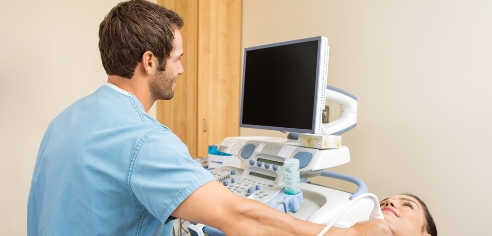
Thyroid ultrasound uses sound waves to produce pictures of the thyroid gland within the neck. It does not use ionizing radiation and is commonly used to evaluate lumps or nodules found during a routine physical or other imaging exam.This procedure requires little to no special preparation. Leave jewelry at home and wear loose, comfortable clothing. You may be asked to wear a gown.
Dr. Snehal Suryawanshi at Insight Imaging & Bone, Joint & Spine Clinic is a top choice. Her 12 years of experience and commitment to quality patient care make her a reliable choice for all your radiology needs. Insight Imging & bone Joint & Spine Clinic is known for offering excellent patient care. The clinic is located centrally in pimpri chinchwad. It stands close to Dange Chowk & Kalewadi phata, which not only makes it convenient for people from the vicinity to consult the doctor but also for those from other neighbourhoods to seek medical guidance.
Procedure:
Preparation: Depending on the specific purpose of the ultrasound, you may be asked to fast for several hours before the procedure. For abdominal ultrasounds, fasting can help improve the visualization of certain organs, like the gallbladder. You may also be asked to drink water and avoid urinating before a pelvic ultrasound to have a full bladder, which helps provide better images of the pelvic organs.
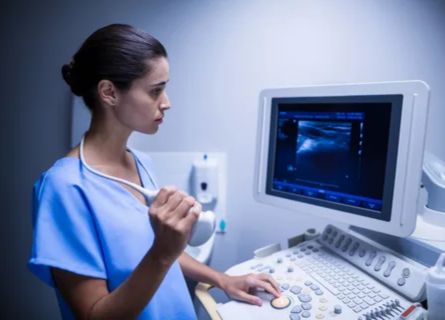
Ultrasound Device: A small handheld device called a transducer is used to conduct the ultrasound examination. This transducer emits high-frequency sound waves, which bounce off the internal structures and are then captured as echoes to create real-time images on a monitor.
Gel Application: A clear gel is applied to the skin over the area to be examined. This gel helps transmit the sound waves and allows the transducer to move smoothly over the skin.
Scanning: The ultrasound technologist or radiologist moves the transducer over the abdomen or pelvic area to capture images of the organs. They may also ask you to change positions or hold your breath briefly to obtain specific views.
Real-Time Imaging: The images are generated in real-time on a computer monitor, allowing the healthcare provider to observe the movement and functionality of the organs as well as any abnormalities.
Uses:
Abdominal and pelvic sonography can be used to evaluate and diagnose a wide range of medical conditions and issues, including:
- Abdominal Organs
- Pelvic Organs
- Abdominal Aorta
- Vascular Assessment
- Evaluation of Abdominal Pain
Why Choose Insight Imaging For Neck/Thyroid?
Selecting Insight Imaging for neck and thyroid sonography, with Dr. Snehal Suryawanshi as a consultant radiologist, offers the advantage of specialized expertise. Dr. Suryawanshi’s extensive experience and the facility’s commitment to advanced technology ensure a comprehensive and precise evaluation of the neck and thyroid. This expertise and access to state-of-the-art equipment can lead to more accurate diagnoses and informed medical decisions, providing confidence in your healthcare experience.. Your health and peace of mind are our top priorities at Insight Imaging. For an Appointment call 8855871212
FAQ
The machine takes your pictures, just like a camera. The advanced machines give far better quality which helps the radiologist and your doctor understand your problem.
The required time depends upon what study needs to performed, cooperation from the patient, and requirement of sedation or contrast.
It depends on what kind of scan is advised to you. Our front office will help you with these queries.
Study of the scan images require lot of dedicated time and expertise of radiologist. We try to report studies in the best possible time.
Latest Blogs / News
Testimonials


