Ovulataion Study
Transthoracic ultrasound (US) of the chest is useful in the evaluation of a wide range of peripheral parenchymal, pleural, and chest wall diseases. Furthermore, it is increasingly used to guide interventional procedures of the chest and pleural space. The technique lends itself to bedside use in the intensive care unit, where suboptimal radiography may mask or mimic clinically significant abnormalities.
Dr. Snehal Suryawanshi at Insight Imaging & Bone, Joint & Spine Clinic is a top choice. Her 12 years of experience and commitment to quality patient care make her a reliable choice for all your radiology needs. Insight Imging & bone Joint & Spine Clinic is known for offering excellent patient care. The clinic is located centrally in pimpri chinchwad. It stands close to Dange Chowk & Kalewadi phata, which not only makes it convenient for people from the vicinity to consult the doctor but also for those from other neighbourhoods to seek medical guidance.
Here's some information about Ovulataion Study Sonography:
Ovulation study sonography, also known as follicular monitoring or ultrasound monitoring, is a medical procedure used to track and monitor the process of ovulation in women. It is commonly employed to assist couples who are trying to conceive, as well as in the management of certain fertility issues.
This imaging allows healthcare providers to monitor the development of ovarian follicles, small sacs that house eggs, and the thickness of the uterine lining. By observing the growth of follicles and hormone levels, such as luteinizing hormone (LH) and estradiol, medical professionals can predict when ovulation is likely to occur. This information aids in determining the optimal timing for intercourse to enhance the chances of conception and is also vital for fertility treatments like intrauterine insemination (IUI) and in vitro fertilization (IVF). Ovulation study sonography is safe, non-invasive, and highly accurate, making it a valuable tool in reproductive medicine for assessing and addressing ovulation-related concerns.
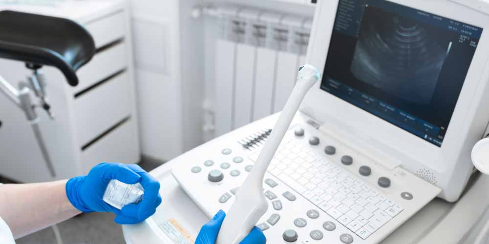
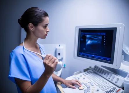
Procedure: During an ovulation study sonography, a transvaginal or transabdominal ultrasound is performed by a trained sonographer or radiologist. A transvaginal ultrasound involves inserting a small probe into the vagina, while a transabdominal ultrasound is performed on the abdominal area. The ultrasound machine uses sound waves to create images of the pelvic organs, including the ovaries and uterus.
FAQ
Ovulation study sonography is a medical imaging procedure that involves using ultrasound to monitor and track the development and release of an egg (ovulation) from the ovaries during a woman’s menstrual cycle.
The machine takes your pictures, just like a camera. The advanced machines give far better quality which helps the radiologist and your doctor understand your problem.
When choosing a facility for ovulation study sonography, such as “Insight Imaging” and consulting with Dr. Snehal Suryawanshi, a Consultant Radiologist, there are several crucial factors to consider. Firstly, verify the credentials and experience of Dr. Suryawanshi and the facility’s staff to ensure they possess the necessary expertise in reproductive and pelvic ultrasound. The quality of equipment and technology is equally vital, as it directly influences the accuracy of ovulation tracking. Additionally, prioritize privacy and comfort, as this can be a sensitive procedure, and consider the convenience of the facility’s location and operating hours, given the need for multiple visits during a menstrual cycle. Seek referrals or recommendations from your gynecologist or reproductive specialist, as their insights can be invaluable.
The required time depends upon what study needs to performed, cooperation from the patient, and requirement of sedation or contrast.
It depends on what kind of scan is advised to you. Our front office will help you with these queries.
Study of the scan images require lot of dedicated time and expertise of radiologist. We try to report studies in the best possible time.
Latest Blogs / News
Testimonials


