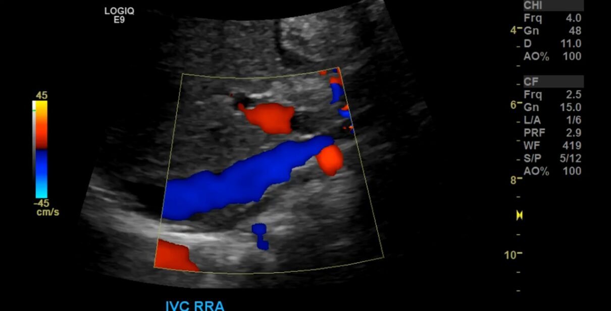
Abdominal Major Vessels Doppler
Abdomen and pelvis sonography, also known as abdominal and pelvic ultrasound, is a medical imaging technique that uses high-frequency sound waves to create detailed images of the structures within the abdominal and pelvic regions of the body. This non-invasive and safe imaging method is commonly used by healthcare providers to evaluate and diagnose a variety of medical conditions affecting the organs in these areas.
Dr. Snehal Suryawanshi at Insight Imaging & Bone, Joint & Spine Clinic is a top choice. Her 10 years of experience and commitment to quality patient care make her a reliable choice for all your radiology needs. Insight Imging & bone Joint & Spine Clinic is known for offering excellent patient care. The clinic is located centrally in pimpri chinchwad. It stands close to Dange Chowk & Kalewadi phata, which not only makes it convenient for people from the vicinity to consult the doctor but also for those from other neighbourhoods to seek medical guidance.
Here's some information about Abdominal Major Vessels Doppler:
Procedure:
Preparation: Depending on the specific purpose of the ultrasound, you may be asked to fast for several hours before the procedure. For abdominal ultrasounds, fasting can help improve the visualization of certain organs, like the gallbladder. You may also be asked to drink water and avoid urinating before a pelvic ultrasound to have a full bladder, which helps provide better images of the pelvic organs.
Ultrasound Device: A small handheld device called a transducer is used to conduct the ultrasound examination. This transducer emits high-frequency sound waves, which bounce off the internal structures and are then captured as echoes to create real-time images on a monitor.
Gel Application: A clear gel is applied to the skin over the area to be examined. This gel helps transmit the sound waves and allows the transducer to move smoothly over the skin.
Scanning: The ultrasound technologist or radiologist moves the transducer over the abdomen or pelvic area to capture images of the organs. They may also ask you to change positions or hold your breath briefly to obtain specific views.
Real-Time Imaging: The images are generated in real-time on a computer monitor, allowing the healthcare provider to observe the movement and functionality of the organs as well as any abnormalities.
Uses:
Abdominal and pelvic sonography can be used to evaluate and diagnose a wide range of medical conditions and issues, including:
- Abdominal Organs
- Pelvic Organs
- Abdominal Aorta
- Vascular Assessment
- Evaluation of Abdominal Pain
Why Choose Insight Imaging For Abdominal Major Vessels Doppler?
Choose Insight Imaging for Abdomen/Pelvis Sonography for the assurance of accuracy and excellence. With Dr. Snehal Suryawanshi, a distinguished Consultant Radiologist, on our team, you’ll benefit from top-tier expertise. Our state-of-the-art technology and patient-centered care make us the ideal choice for precise and comfortable sonography. Your health and peace of mind are our top priorities at Insight Imaging. For an Appointment call 8855871212
FAQ
The machine takes your pictures, just like a camera. The advanced machines give far better quality which helps the radiologist and your doctor understand your problem.
Abdominal Major Vessels Doppler is a diagnostic imaging technique that uses ultrasound to assess blood flow and detect abnormalities in major abdominal blood vessels such as the aorta, renal arteries, and mesenteric arteries.
Insight Imaging is the ideal choice for Abdominal Major Vessels Doppler, bolstered by the presence of Dr. Snehal Suryawanshi, a highly experienced Consultant Radiologist. Their commitment to cutting-edge technology ensures precise results, while a patient-centric approach guarantees a comfortable and efficient experience.
Abdominal Major Vessels Doppler typically takes about 30 to 60 minutes, depending on the complexity of the examination.
It depends on what kind of scan is advised to you. Our front office will help you with these queries.
Study of the scan images require lot of dedicated time and expertise of radiologist. We try to report studies in the best possible time.


