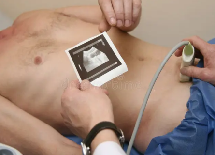
Scrotum sonography, also known as testicular ultrasound, is a non-invasive imaging technique that plays a crucial role in diagnosing various testicular conditions. It utilizes high-frequency sound waves to create detailed images of the scrotum and its contents, including the testicles, epididymis, and surrounding tissues. This procedure is invaluable in assessing a wide range of medical concerns affecting the male reproductive system.
- Detection of Testicular Masses and Tumors
One of the primary benefits of scrotum sonography is its ability to detect testicular masses and tumors. These can include benign conditions like epididymal cysts or potentially serious issues such as testicular cancer. The ultrasound images provide detailed information about the size, location, and characteristics of any abnormal growths, aiding in early diagnosis and treatment planning.
- Evaluation of Testicular Pain
Scrotal pain can be caused by various conditions such as orchitis (inflammation of the testicles), epididymitis (inflammation of the epididymis), or testicular torsion (twisting of the spermatic cord). Scrotum sonography helps in identifying the underlying cause by visualizing inflammation, fluid accumulation, or structural abnormalities within the scrotal sac.
- Assessment of Infertility Issues
Infertility in men can sometimes be linked to anatomical abnormalities within the testicles or epididymis. Scrotum sonography can reveal structural issues such as varicoceles (enlarged veins within the scrotum), which may impair sperm production or flow. By identifying these conditions, healthcare providers can recommend appropriate treatments to improve fertility potential.
- Diagnosis of Testicular Trauma
In cases of trauma to the scrotum, such as sports injuries or accidents, scrotum sonography is crucial for evaluating damage to the testicles and surrounding tissues. It helps in assessing the extent of injury, detecting hematomas (blood clots), or identifying testicular rupture, guiding timely medical interventions.
- Monitoring Testicular Conditions
For patients with known testicular conditions or those undergoing treatment, scrotum sonography serves as a valuable tool for monitoring disease progression or response to therapy. Follow-up ultrasound examinations can track changes in the size and appearance of lesions, providing vital information for ongoing management.
- Non-Invasive and Painless Procedure
One of the significant advantages of scrotum sonography is its non-invasive nature and lack of radiation exposure, making it safe for repeated use as needed. The procedure is generally painless and well-tolerated by patients, involving the application of gel on the scrotal skin followed by the movement of a transducer over the area to capture images.
- Quick and Accurate Results
Scrotum sonography yields rapid results, with images instantly available for interpretation by radiologists or healthcare providers. This facilitates prompt diagnosis and enables timely decision-making regarding further investigations or treatment options.
Scrotum sonography is an indispensable diagnostic tool for evaluating various testicular conditions ranging from benign cysts to potentially life-threatening tumors. Its ability to provide detailed, real-time images of the scrotal anatomy helps in early detection, accurate diagnosis, and effective management of male reproductive health issues. For anyone experiencing scrotal pain, infertility concerns, or other symptoms related to the testicles, consulting a radiologist for scrotum sonography can provide valuable insights and guide appropriate medical care.
Summary:
Scrotum sonography, offered at Insight Imaging Clinic Pune under the expertise of Dr. Snehal Suryawanshi, located in Thergaon, Pune, is pivotal in diagnosing testicular conditions. Known as the ‘Best Radiodiagnosis Clinic in Pune’, Insight Imaging Clinic provides advanced diagnostic services ensuring accurate and timely healthcare solutions.


