Demystifying 2D Echocardiography: A Comprehensive Overview
In the realm of cardiac diagnostics, 2D echocardiography stands as a cornerstone technology. It provides invaluable insights into the structure and function of the heart, aiding in the diagnosis and management of various cardiovascular conditions. In this blog post, we will demystify 2D echocardiography, offering a comprehensive overview of this critical imaging technique, with insights from Dr. Snehal Suryawanshi at Insight Imaging.
Understanding 2D Echocardiography:
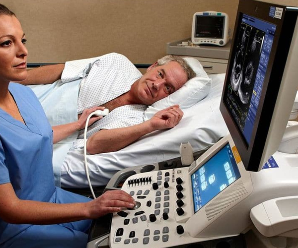
2D echocardiography, also known as two-dimensional echocardiography, is a non-invasive imaging technique that uses high-frequency sound waves (ultrasound) to create detailed, real-time images of the heart. These images are 2D representations of the heart’s chambers, valves, and major blood vessels. The procedure is safe, painless, and does not involve ionizing radiation.
Key Components of 2D Echocardiography:
- Transducer: A handheld device called a transducer emits ultrasound waves and receives the echoes that bounce back from the heart. Different types of transducers are used for various echocardiographic views.
- Echocardiographic Views: During the procedure, the transducer is placed on the patient’s chest or in specific locations to obtain different views of the heart. These views include the parasternal, apical, and subcostal views, each providing unique information about the heart’s anatomy and function.
- Image Formation: The echoes received by the transducer are processed by a computer to create real-time 2D images of the heart. These images can be displayed on a monitor and recorded for further analysis.
Applications of 2D Echocardiography:
2D echocardiography has a wide range of clinical applications, including:
- Assessment of Cardiac Anatomy: It allows for the evaluation of the heart’s size, shape, and overall structure. This is crucial in diagnosing congenital heart defects, valve abnormalities, and other structural heart diseases.
- Evaluation of Cardiac Function: Echocardiography provides insights into the heart’s pumping ability, helping diagnose conditions such as heart failure, cardiomyopathy, and ischemic heart disease.
- Valve Function Assessment: It assesses the function of heart valves, detecting conditions like valve stenosis or regurgitation.
- Doppler Echocardiography: This technique assesses blood flow within the heart and major blood vessels, helping to diagnose conditions like aortic stenosis and mitral regurgitation.
Insight Imaging’s Commitment to 2D Echocardiography:
At Insight Imaging, led by Dr. Snehal Suryawanshi, 2D echocardiography is conducted with the utmost precision and care. Their team of skilled sonographers and cardiologists are trained in obtaining high-quality images and interpreting them accurately. This commitment ensures that patients receive reliable diagnostic information that informs their treatment plans.
Conclusion:
2D echocardiography is a powerful tool in the diagnosis and management of cardiovascular diseases. It provides detailed insights into the heart’s structure and function without the need for invasive procedures. At Insight Imaging, the expertise of professionals like Dr. Snehal Suryawanshi ensures that patients receive the highest standard of care when undergoing 2D echocardiography. This technology continues to play a pivotal role in enhancing our understanding of cardiac health and improving patient outcomes.