USG Thorax
Transthoracic ultrasound (US) of the chest is useful in the evaluation of a wide range of peripheral parenchymal, pleural, and chest wall diseases. Furthermore, it is increasingly used to guide interventional procedures of the chest and pleural space. The technique lends itself to bedside use in the intensive care unit, where suboptimal radiography may mask or mimic clinically significant abnormalities.
Dr. Snehal Suryawanshi at Insight Imaging & Bone, Joint & Spine Clinic is a top choice. Her 12 years of experience and commitment to quality patient care make her a reliable choice for all your radiology needs. Insight Imging & bone Joint & Spine Clinic is known for offering excellent patient care. The clinic is located centrally in pimpri chinchwad. It stands close to Dange Chowk & Kalewadi phata, which not only makes it convenient for people from the vicinity to consult the doctor but also for those from other neighbourhoods to seek medical guidance.
Here's some information about USG Thorax sonography:
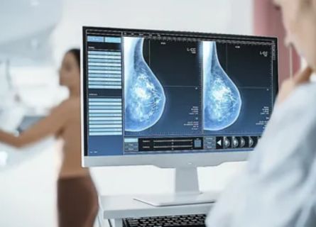
Sonomammography, also known as breast ultrasound, can be performed on one or both breasts depending on the clinical indication and the patient’s specific circumstances.
Purpose: Thorax sonography is used to evaluate and diagnose various medical conditions affecting the chest, including the heart, lungs, pleura, and mediastinum (the area between the lungs). It can help identify abnormalities such as fluid accumulation, masses, cysts, or other structural anomalies.
Preparation: Typically, there is minimal preparation required for a thorax sonography. Patients may be asked to remove clothing from the upper body and wear a gown. It’s important to inform the healthcare provider about any relevant medical history or allergies.
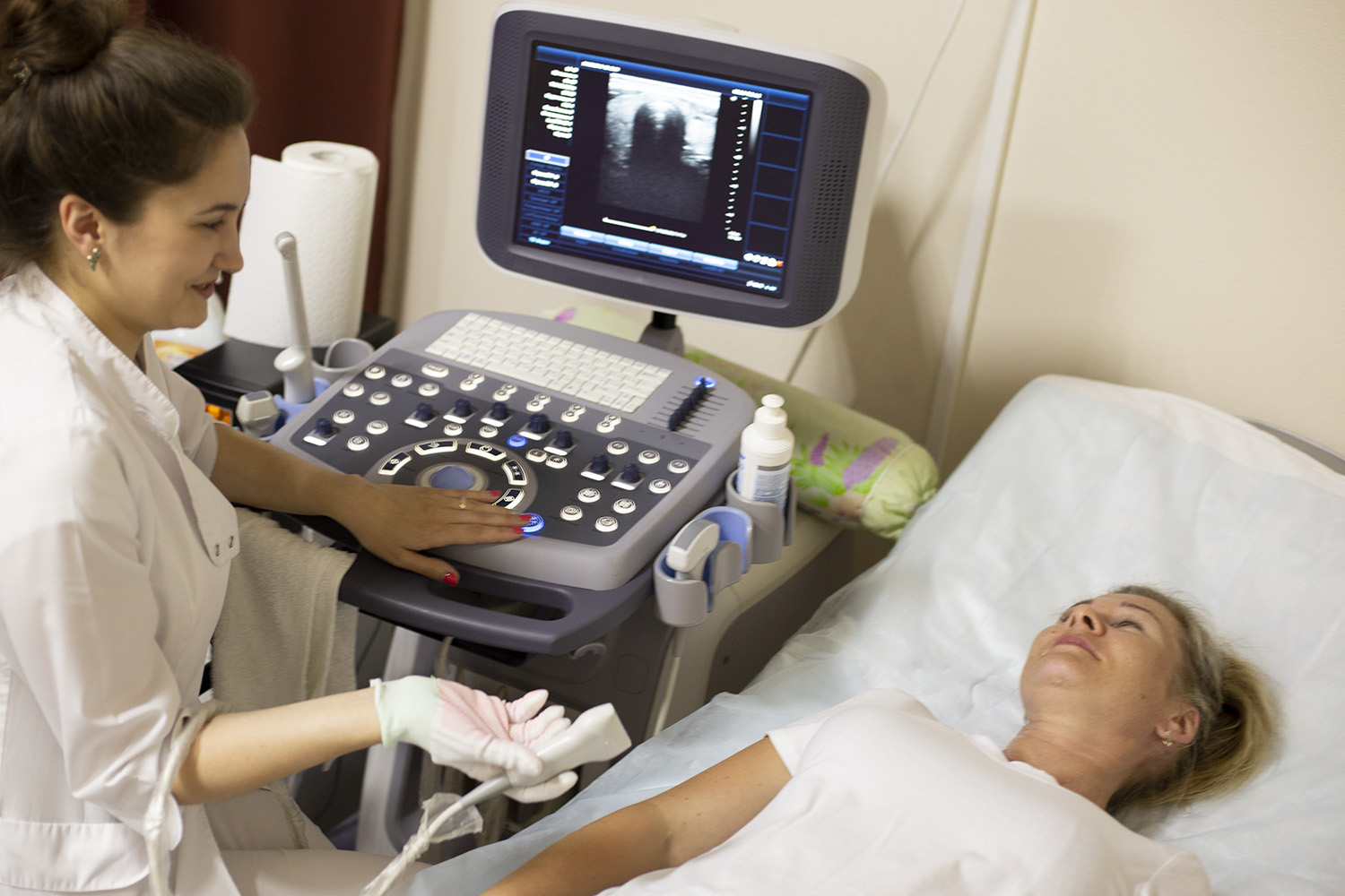
Procedure: During the procedure, a gel is applied to the skin over the chest area to help transmit the sound waves. A transducer, which emits and receives sound waves, is then moved over the chest. The sound waves bounce off the internal structures, and the returning echoes are used to create real-time images on a monitor.
Types of Thorax Sonography:
- Cardiac Sonography: This focuses on the heart and is used to assess its structure and function. It can help diagnose conditions such as heart disease, valve problems, or pericardial effusion (fluid around the heart.
- Pulmonary Sonography: This examines the lungs and can detect conditions like pleural effusion (fluid around the lungs) or pneumothorax (collapsed lung).
- Mediastinal Sonography: It evaluates the mediastinum, which contains structures like the thymus, lymph nodes, and major blood vessels.
Uses:
Thorax sonography, also known as thoracic ultrasound or chest ultrasound, is a medical imaging technique that utilizes high-frequency sound waves to visualize and assess structures within the chest cavity. It has a wide range of clinical uses, making it a valuable tool for healthcare professionals in various specialties.
Thorax sonography is a versatile imaging technique with uses ranging from diagnosing pulmonary and pleural conditions to assessing cardiac health. Its non-invasive nature and ability to provide real-time imaging make it an essential tool in modern medicine.
FAQ
Dr. Snehal Suryawanshi’s role as a Consultant Radiologist at Insight Imaging adds a significant level of credibility to the practice. Her dedication to the field and years of specialized practice ensure that patients receive the highest quality care and the most accurate diagnoses. With thorax sonography being a critical diagnostic tool for various pulmonary and chest-related conditions, having a highly qualified expert like Dr. Suryawanshi can make all the difference in the accuracy and effectiveness of the procedure. Your health and peace of mind are our top priorities at Insight Imaging. For an Appointment call 8855871212
Explanation of thorax sonography as a medical imaging technique.
The machine takes your pictures, just like a camera. The advanced machines give far better quality which helps the radiologist and your doctor understand your problem.
A thorax sonography is usually a relatively quick procedure, often taking less than 30 minutes to complete.
It depends on what kind of scan is advised to you. Our front office will help you with these queries.
Study of the scan images require lot of dedicated time and expertise of radiologist. We try to report studies in the best possible time.
Latest Blogs / News
Testimonials


