Pregnancy is a journey filled with anticipation, excitement, and many questions about the health and development of the baby. Abdomen sonography, also known as an abdominal ultrasound, is a critical tool used by healthcare professionals to monitor the fetus’s growth and development throughout pregnancy. This non-invasive procedure provides invaluable insights, ensuring both the mother’s and baby’s well-being.
What is Abdomen Sonography?
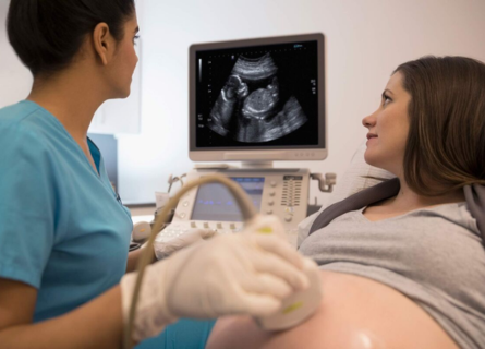
Abdomen sonography is an imaging technique that uses high-frequency sound waves to create images of the organs and structures inside the abdomen. During pregnancy, this method is employed to visualize the developing fetus, assess its growth, and detect any potential abnormalities.
How Does Abdomen Sonography Work?
The procedure is straightforward and safe. A gel is applied to the pregnant woman’s abdomen, and a handheld device called a transducer is moved over the skin. The transducer emits sound waves that bounce off the baby’s body and are captured back to create an image on a monitor. These images help the doctor to evaluate the fetus’s health and development.
Importance of Abdomen Sonography in Pregnancy
- Confirming Pregnancy: The first ultrasound is usually performed around 6-8 weeks of pregnancy to confirm the pregnancy and establish a due date. It ensures the pregnancy is developing inside the uterus and rules out ectopic pregnancies.
- Monitoring Fetal Development: Regular ultrasounds track the growth and development of the fetus. They measure the baby’s size, estimate weight, and check for normal development patterns. This helps in identifying growth restrictions or overgrowth, which might indicate underlying health issues.
- Detecting Congenital Abnormalities: Detailed anatomy scans, typically performed around 18-22 weeks, can detect structural abnormalities in the fetus. This early detection is crucial for planning necessary medical or surgical interventions after birth.
- Assessing Fetal Position: As the pregnancy progresses, ultrasound helps in determining the baby’s position. This aspect is crucial for determining the most effective method of delivery. For instance, if the baby is in a breech position (feet or buttocks first), a cesarean section might be recommended.
- Monitoring Amniotic Fluid Levels: Adequate levels of amniotic fluid are essential for the baby’s development. Ultrasound helps measure the fluid levels, ensuring they are within a healthy range. Both too much and too little amniotic fluid can cause complications.
- Checking Placental Health: The placenta provides oxygen and nutrients to the growing baby. Ultrasound evaluates the placental position and health, identifying issues like placenta previa (placenta covering the cervix) or placental abruption (placenta detaching from the uterus wall).
- Observing Fetal Movements and Heartbeat: Ultrasound allows for monitoring of the baby’s movements and heartbeat, reassuring parents and doctors of the fetus’s well-being.
- Diagnosing Multiple Pregnancies: If a woman is carrying more than one baby, ultrasound can confirm multiple pregnancies and help monitor the health and development of each fetus.
Types of Abdomen Sonography in Pregnancy
- Transabdominal Ultrasound: The most common type where the transducer is moved across the abdomen.
- Transvaginal Ultrasound: Used in early pregnancy or when a clearer image is needed, where the transducer is inserted into the vagina.
- 3D and 4D Ultrasounds: These advanced ultrasounds provide three-dimensional images and real-time video of the fetus, offering more detailed visualization.
Preparing for an Abdomen Sonography
Preparation for an abdomen sonography is minimal but important for clear images. Pregnant women are usually advised to drink plenty of water before the scan to fill the bladder, which helps in obtaining better images of the fetus.
Safety and Benefits
Abdomen sonography is a safe procedure with no known risks to the mother or the baby. It does not involve radiation, making it a preferred method for monitoring fetal development. The benefits include early detection of potential issues, reassurance for the parents, and crucial information for the healthcare team to provide the best possible care.
Insight Imaging Clinic in Thergaon, Pune
Insight Imaging Clinic in Thergaon, Pune, is renowned for its state-of-the-art facilities and expert radiologists. The clinic offers comprehensive abdomen sonography services, ensuring accurate monitoring of fetal development. With advanced imaging technology and a patient-centric approach, Insight Imaging Clinic is dedicated to providing the highest standard of care for expecting mothers.
Summary
Abdomen sonography is essential for monitoring fetal development throughout pregnancy. It confirms pregnancy, tracks growth, detects abnormalities, and assesses fetal health. Insight Imaging Clinic in Thergaon, Pune, offers advanced abdomen sonography services, ensuring comprehensive care for expecting mothers. For expecting parents in Pune, Dr. Snehal Suryawanshi and Insight Imaging Clinic provide reliable and accurate ultrasound services, making them the best choice for prenatal care. With their expertise and advanced technology, they ensure the well-being of both mother and baby. Visit the Best Radiodiagnosis Clinic in Pune for expert care and peace of mind during your pregnancy journey.