


The Gentle Beam: Why Digital Precision is the New Safety Standard for Pediatric X-Rays
Rohan stood in the hallway of a busy orthopedic center in Wakad, watching his seven-year-old daughter, Ishani, climb onto the cold, metal X-ray table. She had taken a nasty tumble during her skating lessons at Balewadi Stadium, and her wrist was swelling rapidly. As the technician pulled a heavy lead apron over Rohan, a wave of anxiety hit him.
“Is she getting too much radiation?” he wondered. “She’s so small. Her bones are still growing. I’ve heard these scans can be risky for kids, but how else will we know if it’s broken?”
Rohan’s fear is one shared by thousands of parents across Pune. In the past, getting an X-ray meant exposing a child to a significant “blast” of radiation because older, film-based machines required higher doses to produce a clear image. But medicine has evolved. At Insight Imaging & Bone, Joint & Spine Clinic, we believe that a child’s diagnostic journey should be defined by precision, not “dosage.” By switching to advanced Digital X-Ray technology, we have eliminated the unnecessary risks associated with traditional, high-radiation scans.
The Pediatric Vulnerability: Why “Old” X-Rays are Risky
Children are not just small adults; biologically, they are much more sensitive to radiation. Their cells are dividing rapidly as they grow, which makes their DNA more susceptible to the ionizing effects of X-rays. Furthermore, because children have a longer life expectancy ahead of them, any potential long-term effects of radiation exposure have more time to manifest.
Traditional film X-rays often suffer from “exposure guesswork.” If the dose was slightly too low, the image would be blurry or dark, forcing the technician to repeat the scan—doubling the radiation dose for the child. In a growing body, especially near sensitive areas like the growth plates in the joints, these repeated “hits” of radiation are a risk that modern technology can now avoid.
The Digital Advantage: Speed, Safety, and Clarity
Digital X-Ray technology has revolutionized pediatric diagnostics by applying the same principles that turned bulky film cameras into high-definition smartphones. At Insight Clinic, we utilize Digital Radiography (DR), which offers three life-changing advantages for your child:
The “Low-Dose” Priority: Digital sensors are significantly more sensitive than old-fashioned film. This means we can capture a crystal-clear image using up to 80% less radiation than traditional methods. For a child like Ishani, this means the “exposure” is kept to the absolute biological minimum.
Instant Results, Zero Repeats: With digital technology, the image appears on the doctor’s screen in under three seconds. We no longer have to wait for “film development” only to realize the child moved. If a child fidgets, we know instantly and can adjust, ensuring we get the perfect diagnostic shot the first time.
Post-Processing Power: Unlike a physical film, a digital image can be zoomed, brightened, or contrasted after the scan is taken. If Dr. Snehal Suryawanshi needs to see a tiny hairline fracture in a small wrist bone, she can enhance the digital image on her monitor without ever having to re-expose the child to more radiation.
The “Silent Whispers” of Childhood Bone Injuries
While you wait for a scan, your child’s body is already signaling the severity of the injury. Parents should look for “Point Tenderness”—if the child can point to one exact spot that hurts significantly more than the surrounding area, it is a high indicator of a fracture.
Another subtle sign is “Functional Guarding.” If your child is moving their arm but refuses to use their hand to grip a toy or pick up a snack, the bone might be compromised even if the swelling is minimal. In Pune’s competitive sports culture, we often see children “braving the pain,” but any persistent limp or refusal to bear weight should be met with the safety of a low-dose digital scan rather than a “wait and watch” approach.
The Roadmap to a Safe Recovery: Recommendations
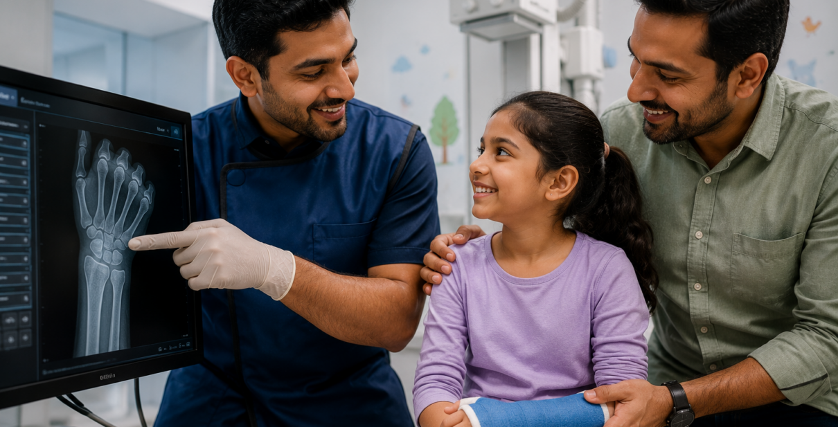
Ishani’s digital X-ray showed a “Greenstick” fracture—a common pediatric injury where the bone bends and cracks but doesn’t break completely. Because the scan was digital, Dr. Snehal was able to provide the orthopedic team with a high-definition map of the growth plate to ensure the injury wouldn’t affect her future height.
- Demand Digital: When your pediatrician recommends an X-ray, specifically ask for a “Digital DR Scan.” It is the most effective way to ensure your child receives the lowest possible radiation dose.
- Lead Shielding is Non-Negotiable: Even with low-dose digital scans, always ensure the clinic provides lead aprons or “gonad shields” to protect the child’s most sensitive organs from scatter radiation.
- Keep a Digital History: One of the best features of digital X-rays is that they are stored as files, not physical films that fade or get lost. At Insight Clinic, we provide digital copies that you can share instantly with specialists, preventing the need for “repeat scans” at different hospitals.
The Insight Solution: Expert Interpretation Meets Safe Tech
At Insight Imaging & Bone, Joint & Spine Clinic, we don’t just provide the technology; we provide the expertise of Dr. Snehal Suryawanshi (MBBS, DNB Radiologist). With 12 years of experience, Dr. Snehal understands that interpreting a child’s X-ray requires a deep knowledge of developing anatomy. What might look like a fracture to an untrained eye could simply be a normal growth plate.
Our Thergaon center is designed to be child-friendly, reducing the “white coat anxiety” that often leads to the movement and blurry images that cause repeat exposures. We combine the world’s safest digital hardware with a human touch that puts both the parent and the child at ease.
Prioritize Your Child’s Safety Today
Insight Imaging & Bone, Joint & Spine Clinic Expert: Dr. Snehal Suryawanshi (Radiologist)
Location: 107, B-Square, Barne Corner, Opposite Atithi Pure Veg, Thergaon, Pune.
Appointments: 8855871212 / 09511763474
Top Services: Digital X-Ray, Pediatric Ultrasound, Bone Density Checks.