Most imaging reports follow a standardized format. Knowing this structure can help you focus on the most important sections:
- Patient Information: Name, age, gender, and date of the test
- Clinical History: Reason for the imaging procedure and any symptoms
- Technique/Procedure: Type of imaging performed
- Findings: Observations made by the radiologist
- Impression/Conclusion: Summarizes the key findings and clinical significance
Focusing on the Findings and Impression sections helps you understand the relevance of the results to your healthcare situation.
Common Terms Used in Diagnostic Reports
Hypoechoic / Hyperechoic (Ultrasound)
- Hypoechoic: Appears darker; may indicate fluid-filled structures or tissue
- Hyperechoic: Appears brighter; may reflect denser or calcified tissue
Attenuation (CT Scan)
Indicates how much X-ray energy is absorbed by tissues:
- Low attenuation: May indicate fluid or fat
- High attenuation: May indicate bone
Signal Intensity (MRI Reports)
- Hyperintense: Bright areas indicating inflammation or tissue changes
- Hypointense: Darker areas, possibly scar tissue, calcifications, or less active lesions
Lesion / Mass / Nodule
Indicates a potentially abnormal area that may be benign or requires further evaluation.
Normal / No Significant Findings
Indicates that the examined area appears healthy.
Tips for Reading Your Imaging Report
- Focus on the Impression Section: Provides an overview of key findings and recommendations.
- Compare Findings with Clinical History: Understanding why the imaging was done helps interpret results.
- Look for Recommendations: Follow-up scans or referrals may be suggested.
- Don’t Panic Over Medical Jargon: Many terms describe appearance, size, or density, not severity.
- Ask Questions: Clarify any doubts with your physician or radiologist. Insight Clinic encourages patient discussions.
Differences Between Ultrasound, CT, and MRI Reports
Ultrasound Reports
- Focus on soft tissues and fluid-filled structures
- Terms: hypoechoic, hyperechoic, cyst
- Often provide real-time functional information, like blood flow
CT Reports
- Offer detailed views of bones, organs, and blood vessels
- Terms: attenuation, calcification, mass density
- Useful in trauma or emergency situations
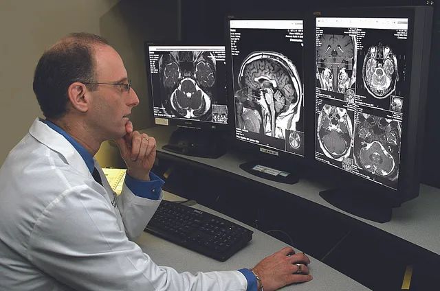
MRI Reports
- Excellent soft tissue contrast
- Terms: hyperintense, hypointense, lesion characteristics
- Commonly used for brain, spine, joints, and musculoskeletal studies
Understanding these differences helps interpret reports from different imaging modalities.
When to Seek Clarification
Consult your doctor or radiologist if:
- You encounter unfamiliar or concerning terms
- Follow-up scans or treatments are recommended
- You want to understand how results relate to your symptoms
At Insight Clinic, patients receive a full explanation of imaging results from experienced staff led by Dr. Snehal Suryawanshi.
The Role of Insight Clinic in Interpreting Reports
Located in Thergaon, Pimpri-Chinchwad, Insight Clinic bridges the gap between technical imaging reports and patient understanding. Services include:
- Ultrasound
- CT scans
- MRI
- Radiologist consultations
The clinic ensures patients receive appropriate imaging and clear instructions to make the process stress-free and informative.
Final Thoughts
Reading diagnostic imaging reports doesn’t have to be confusing. Key points to remember:
- Focus on the Impression/Conclusion section
- Understand the differences between ultrasound, CT, and MRI reports
- Don’t worry about medical jargon—it usually describes appearance, not severity
- Ask questions to clarify findings
With the guidance of Dr. Snehal Suryawanshi at Insight Clinic, patients in Pimpri-Chinchwad can confidently interpret their imaging results and make informed healthcare decisions.