2D Echo Test: What It Detects, Procedure & Who Needs It
When it comes to heart health, many people ignore early warning signs like chest discomfort, breathlessness, palpitations, or unusual fatigue. The truth is: your heart can have structural or functional issues even if your ECG looks normal. This is where a 2D Echo test becomes extremely useful.
A 2D Echocardiography (2D Echo) is a simple, painless test that creates real-time moving images of your heart using ultrasound waves. It helps doctors see how well your heart is functioning—how the valves are working, how strongly the heart muscles are pumping, and whether blood flow is normal.
Let’s break it down clearly: what a 2D Echo detects, how the procedure works, and who should get this test.
What Exactly is a 2D Echo Test?
A 2D Echo is an ultrasound-based test that shows the heart’s structure and motion in real time. “2D” means the images appear in two dimensions, allowing the doctor to visualize the chambers, walls, and valves.
Unlike an ECG (which checks electrical signals), a 2D Echo checks the physical pumping action of the heart. So even if your ECG is normal, your 2D Echo can still reveal serious conditions like weak heart pumping, valve leakage, or fluid around the heart.
What Does a 2D Echo Detect?
A 2D Echo is one of the most informative heart tests because it shows both structure and movement. Here are the major things it can detect:
1) Heart pumping strength (Ejection Fraction)
Your heart’s pumping efficiency is measured using Ejection Fraction (EF). If EF is low, it may suggest heart failure, weak heart muscle, or damage after a heart attack.
2) Heart valve problems
Valves control blood flow direction. A 2D Echo can detect:
- Valve leakage (regurgitation)
- Valve narrowing (stenosis)
- Valve thickening or deformity
Valve issues are common and often silently worsen over time—2D Echo catches them early.
3) Enlarged heart chambers
High BP, valve diseases, and heart failure can cause enlargement of heart chambers. Echo identifies these changes and helps determine severity.
4) Congenital heart defects
Some people are born with small holes or abnormal connections in the heart. 2D Echo can identify congenital issues like ASD, VSD, and other abnormalities.
5) Fluid around the heart (Pericardial effusion)
If fluid collects around the heart due to infection, inflammation, or other causes, echo detects it clearly.
6) Blood clots or masses inside the heart
In certain high-risk cases, a 2D Echo can detect clots, abnormal growths, or infections affecting heart tissues.
7) Pulmonary artery pressure (indirect clue)
Echo may also suggest pulmonary hypertension, which can cause breathlessness and fatigue.
2D Echo vs ECG: What’s the Difference?
Many patients confuse these two tests. Here is the simplest difference:
- ECG = checks heart rhythm & electrical activity
- 2D Echo = checks heart structure & pumping function
So if your symptoms are physical—like breathlessness, swelling, or weakness—2D Echo is more revealing than ECG alone.
Who Needs a 2D Echo Test?
You should consider a 2D Echo if you fall into any of these categories:
1) If you have symptoms like:
- Chest pain or heaviness
- Breathlessness (especially on walking or climbing stairs)
- Palpitations (fast/irregular heartbeat)
- Swelling in legs/ankles
- Dizziness, fainting, or unexplained weakness
Don’t assume “it’s only gas” or “it’s stress.” That mindset is dangerous.
2) If you have high blood pressure (BP)
Long-term BP affects the heart muscle and can silently cause thickening of the heart wall. A 2D Echo is one of the best tests to monitor heart impact from hypertension.
3) If you have diabetes or high cholesterol
These conditions increase risk of heart disease. Even without symptoms, doctors often recommend 2D Echo for better screening.
4) If you have had a heart attack or stent
After any cardiac event, a 2D Echo helps evaluate:
- damage level
- pumping efficiency (EF)
- recovery progress
5) If your ECG is abnormal
ECG changes can be a signal of heart stress, rhythm disorders, or heart enlargement. Echo confirms the structural cause behind ECG changes.
6) If you have a heart murmur
A murmur often suggests valve issues. A 2D Echo is the correct test to confirm valve health.
7) If your family has a history of heart disease
If heart disease runs in your family, early screening gives you an advantage. Prevention is always cheaper than treatment.
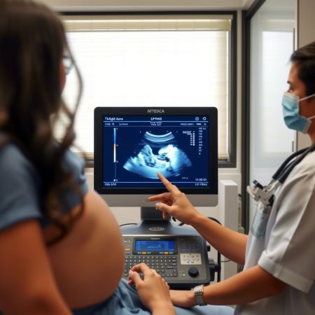
2D Echo Procedure: What Happens During the Test?
A 2D Echo is simple and usually takes 15 to 30 minutes.
Step-by-step process:
- You’ll be asked to lie down on a bed, usually on your left side.
- A gel is applied to the chest area (to help ultrasound waves travel).
- The technician/doctor moves a probe (transducer) over the chest.
- The monitor shows a live moving image of your heart.
- Images and measurements are recorded for diagnosis.
No injections. No pain. No radiation.
Preparation Before 2D Echo
Good news: in most cases, no special preparation is needed.
Still, follow these basic tips:
- Wear comfortable clothes
- Carry previous heart reports (ECG, BP record, lab reports)
- Avoid heavy meals just before the test (optional but helpful)
Your doctor may guide differently if you are undergoing a specific type like stress echo (rare for basic clinic screening).
Is 2D Echo Safe?
Yes. A 2D Echo uses ultrasound waves, not X-rays. It is completely safe for:
- elderly patients
- pregnant women
- repeated follow-ups
- children
Conclusion
A 2D Echo test is not just for people with major heart issues. It’s for anyone who wants clarity about their heart’s pumping strength and valve function. If you’re facing symptoms like breathlessness, palpitations, swelling, or chest discomfort, delaying diagnosis is a bad decision. A timely 2D Echo can detect problems early and guide the right treatment before complications occur.
Doctor: Dr. Snehal Suryawanshi
Clinic Address: 107, B-Square, Barne Corner, Opposite Hotel Atithi Pure Veg, Thergaon, Pune, Maharashtra – 411033
Website: https://insightclinicpune.com/