When you hear the word sonography, you may immediately think of pregnancy scans. But sonography (also called ultrasound) is much more than that. It is one of the safest and most commonly used diagnostic tests in modern healthcare. Doctors use it to look inside the body in real time, without surgery, radiation, or long recovery time.
A sonography test helps detect many health conditions early, allowing doctors to start treatment sooner and avoid complications. Whether it’s abdominal pain, swelling, thyroid issues, kidney stones, or pregnancy monitoring—sonography plays a crucial role in accurate diagnosis.
What Is a Sonography Test?
Sonography is a medical imaging technique that uses high-frequency sound waves to create images of the inside of your body. Unlike X-rays or CT scans, ultrasound does not use radiation.
A special handheld device called a transducer is moved over the skin after applying gel. The sound waves travel into the body and bounce back from organs, tissues, and fluids. These reflected waves are converted into images on a screen.
Because it provides live images, sonography is very useful for checking movement, blood flow, and organ function.
Why Is Sonography Done?
Doctors recommend sonography for several reasons, but the main purpose is simple: to find out what’s happening inside the body when symptoms are unclear. Many health conditions do not show symptoms early. Sonography helps detect issues before they become serious.
Some common reasons include:
- Pain in the abdomen or pelvis
- Swelling in any part of the body
- Suspected kidney stones or gallstones
- Thyroid swelling or lumps
- Liver disorders like fatty liver
- Uterine fibroids, ovarian cysts, or PCOD
- Pregnancy checks and fetal development monitoring
- Checking fluid collection, abscess, or infection
- Evaluating organs such as kidney, liver, pancreas, bladder, and spleen
In short, sonography helps doctors get a clear picture quickly and safely.
How Sonography Helps in Diagnosis
Sonography is not just a “scan.” It is a powerful diagnostic tool that supports doctors in making accurate decisions. Here’s how it helps:
1) Early detection of disease
Many conditions like cysts, tumors, stones, or inflammation can be detected at an early stage through sonography. Early diagnosis means earlier treatment, better outcomes, and fewer complications.
2) Identifying the cause of pain
Abdominal pain can have multiple causes—gas, infection, liver issues, kidney stones, gallbladder problems, appendicitis, or gynecological conditions. Sonography helps narrow down the exact reason without delay.
3) Evaluating organ structure
Ultrasound can show size, shape, and structure of organs. If the liver is enlarged, kidneys are swollen, or there is fluid around organs, sonography will detect it clearly.
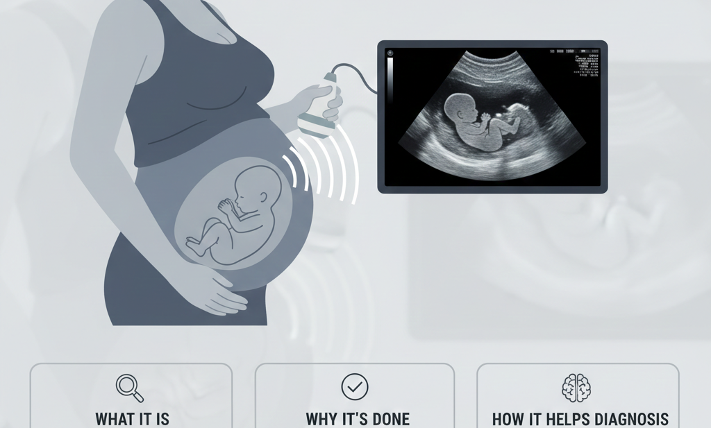
4) Checking pregnancy health
In pregnancy, sonography is used to confirm:
- pregnancy location (important for detecting ectopic pregnancy)
- fetal heartbeat
- growth development
- placenta position
- amniotic fluid levels
- anomalies or structural problems in later months
This makes sonography essential for both mother and baby’s safety.
5) Guiding further treatment
Sometimes sonography findings help doctors decide the next step:
- medication management
- surgery
- further tests like CT or MRI
- specialist referral
So, ultrasound is often the first and most important step in the diagnostic plan.
Common Types of Sonography Tests
Sonography is done for different body parts depending on symptoms. Some common types are:
1) Abdominal Ultrasound
Checks organs like liver, gallbladder, pancreas, spleen, and kidneys. Helps diagnose:
- fatty liver
- gallstones
- liver infection
- kidney stones
- abdominal swelling
2) Pelvic Ultrasound
Used for reproductive organs. Helps detect:
- fibroids
- ovarian cysts
- PCOD/PCOS
- uterine abnormalities
- pelvic infection
3) Thyroid Ultrasound
Used for neck swelling, thyroid nodules, and thyroid gland evaluation.
4) Obstetric Ultrasound
Pregnancy ultrasound done in early pregnancy, NT scan, anomaly scan, and growth scans.
5) Soft Tissue / Lump Ultrasound
Used to evaluate swelling, abscess, or cyst in different parts of the body.
Preparation for Sonography (Important!)
Preparation depends on the type of ultrasound. If preparation is not correct, the scan image may not be clear.
- For abdominal ultrasound: You may need 6–8 hours fasting
- For pelvic ultrasound: You may need a full bladder (drink water before scan)
- For pregnancy scan: depends on stage; early pregnancy may need full bladder
- For thyroid/soft tissue ultrasound: usually no special preparation needed
Always follow the clinic’s instructions for best accuracy.
What Happens During the Procedure?
A typical sonography test is simple and painless.
- You lie down comfortably
- Gel is applied on the area
- The transducer is moved over the skin
- Images appear on the screen
- The doctor observes and captures important images
- The gel is wiped off after completion
The test usually takes 10 to 30 minutes depending on complexity.
Is Sonography Safe?
Yes—sonography is considered extremely safe. Since it uses sound waves and not radiation, it can be repeated multiple times without risk. This is one of the biggest reasons it is commonly used for pregnancy monitoring as well.
Conclusion
A sonography test is one of the most important and reliable diagnostic tools available today. It is safe, painless, and gives quick results. Whether you are facing unexplained pain, swelling, thyroid issues, digestive problems, kidney stones, or pregnancy monitoring—sonography provides valuable information that helps doctors diagnose problems early and accurately.
If you have symptoms that should not be ignored, getting a timely sonography test can help detect the issue early and guide the right treatment.
Clinic Name: Insight Imaging Clinic Pune
Doctor Name: Dr. Snehal Suryawanshi (Radiologist)
Address: 107, B-Square, Barne Corner, Opposite Hotel Atithi Pure Veg, Thergaon, Pune, Maharashtra – 411033